
26 Jul Zone II Flexor Tendon Repair: the patient guide
Zone 2 flexor tendon injury is one of the most demanding problems in hand surgery.
This guide covers the anatomy, surgical principles, and rehabilitation pathway that governs recovery after a zone 2 repair, written for patients preparing for surgery and for clinicians building or reviewing their protocols.
What is Zone 2?
Zone 2 sits in the narrow fibro-osseous tunnel between the A1 pulley and the insertion of the flexor digitorum superficialis, a region historically called no man’s land because outcomes were so unpredictable. Modern surgical technique and structured rehabilitation have changed that, but the rehabilitation window remains tight and the consequences of getting it wrong are significant.
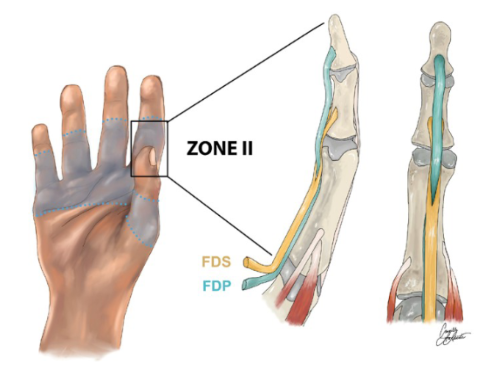
Illustration credit: Research Gate
Why Zone 2 is different
The flexor tendons of the fingers run inside a pulley system that keeps them pressed close to bone. Zone 2 spans from the first annular (A1) pulley at the base of the finger to just past the second joint (PIP joint), where the flexor digitorum superficialis (FDS) splits and inserts. Both the FDS and flexor digitorum profundus (FDP) run through this zone, meaning a laceration here often involves both tendons.
The problem is space. The tendons glide within a sheath that has almost no tolerance for swelling, adhesion, or bulk. Any repair adds volume. Any inflammatory response adds more. The result, without careful management, is a tendon that heals to its surrounding sheath rather than gliding freely through it, producing a stiff finger that lacks active flexion even though the repair itself is intact.
This is the core challenge of zone 2 rehabilitation: protecting the repair from rupture while simultaneously preventing adhesion formation.
What happens in surgery?
A zone 2 repair is typically performed under regional anaesthesia, often with the patient awake so the surgeon can ask them to move the finger and assess tendon glide intraoperatively. The tendons are retrieved, trimmed, and repaired using a core suture technique, most commonly a four- or six-strand configuration, combined with a circumferential epitendinous suture to smooth the repair site and increase resistance to gap formation.
Core strand number matters. Biomechanical studies consistently show that four-strand repairs withstand significantly more force than two-strand repairs before gap formation occurs. Six-strand repairs are stronger still, though technically more demanding. The repair strength is what determines how early and how aggressively rehabilitation can begin.
Rehabiliation Protocols
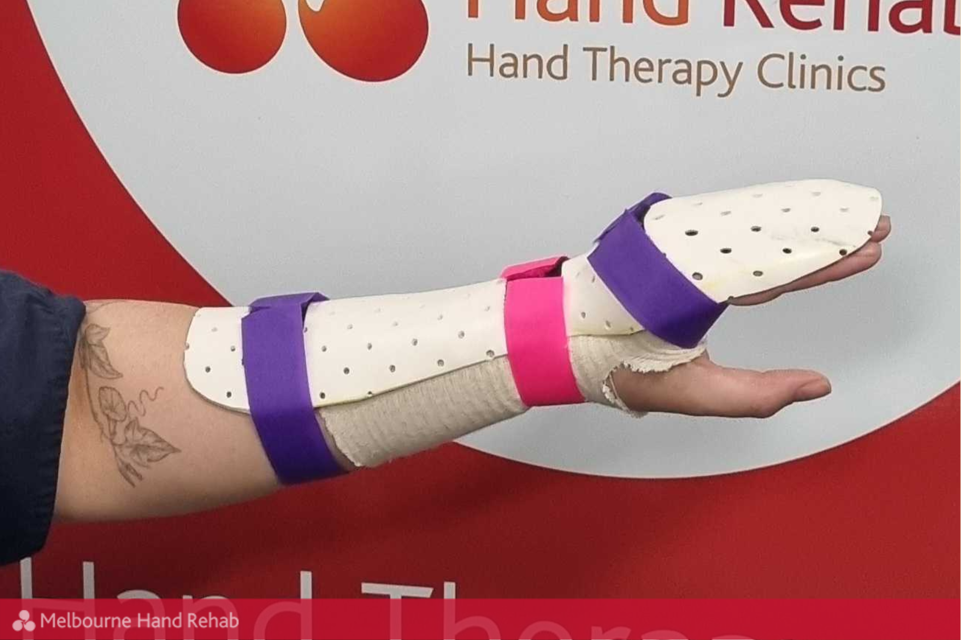
Rehabilitation after zone 2 repair is delivered in close partnership between the operating surgeon and a hand therapist. The goal across the first twelve weeks is to achieve a balance between tendon excursion (movement) and protection from tensile load. Early active range of motion, introduced within the protected environment of the dorsal blocking splint, is central to modern zone 2 management. Moving the finger early prevents adhesion formation without compromising the repair, provided the geometry of the splint is maintained.
Weeks 0 to 2: Dorsal Blocking Splint and Early Active Motion
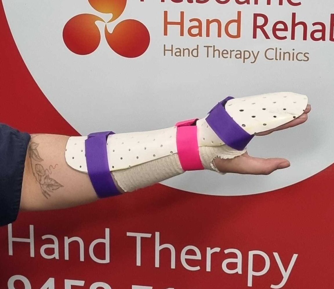
Immediately post-repair, the hand is placed in a dorsal blocking splint with the wrist in approximately 20 to 30 degrees of flexion, the metacarpophalangeal (MCP) joints at 60 to 70 degrees of flexion, and the interphalangeal (IP) joints at full extension. Holding the wrist and MCP joints in flexion slackens the flexor tendons proximally, so any movement of the fingers within that range generates tendon glide without putting the repair under meaningful tensile load. The finger travels through a useful arc while the force across the repair site stays well below the threshold at which sutures gap or fail. Movement starts within days of surgery because the geometry of the splint makes it safe to do so.
Within the first 48 to 72 hours, the patient begins gentle, relaxed range of motion exercises within the hood of the splint under therapist supervision. The exercises are slow and unpressured. The finger moves through whatever arc is available without pushing into resistance or end-range tension. The splint limits how far the finger can extend, which means the tendon is never placed under stretch during this phase. Repetitions are low and spread across the day rather than concentrated into a single session.
Weeks 2 to 4: Increasing Excursion
As wound healing progresses, the therapy focus shifts to maximising differential glide between the FDS and FDP and preventing early adhesion. Creating a hook fist (MCP joints extended, IP joints flexed) isolates FDP excursion. Creating a composite fist (all joints into composite flexion with IP joints leading) generates maximum tendon travel. Both exercises are performed as gentle repetitions within the protected splint, with the wrist maintained in flexion to reduce tensile load.
Oedema management continues through this period with retrograde massage, compressive wrapping, and elevation. Scar massage begins once the wound is fully closed.
Weeks 4 to 6: Progressive Active Flexion
At the four-week mark, with a well-healed repair, the protocol advances to active unresisted flexion through a wider range. The dorsal blocking splint is gradually extended toward neutral wrist, and place-and-hold exercises progress to full active composite fisting. Some protocols introduce active extension blocking at this stage to encourage FDS-FDP differential glide.
The patient is typically still in the splint for protection between exercises. Night splinting continues.
Weeks 6 to 8: Splint Weaning and Functional Use
The dorsal blocking splint is discontinued progressively. The patient begins using the hand for light functional tasks, activities of daily living that do not involve gripping, lifting, or resisted pinch. Joint mobilisation to the PIP joint is introduced if extension lag or flexion deficit has developed.
Weeks 8 to 12: Strengthening
Gentle progressive resistance begins after week eight, starting with putty or therapeutic foam at low resistance and advancing based on tendon palpation and patient tolerance. The repair is mature enough at this stage to tolerate strengthening loads, provided there is no evidence of gap formation, usually assessed clinically by the presence of a tendon bowstring or sudden loss of active flexion range.
Return to manual work, sport, or gripping activities is generally not permitted until twelve weeks at the earliest and is often extended to four to six months depending on the demands of the activity.
Complications to Know
Rupture is the most feared early complication, most common between weeks two and four when the repair has lost its initial suture strength but has not yet developed biological healing. Patients and treating therapists need to recognise sudden loss of active flexion and seek urgent review.
Adhesion formation is the most common late complication, producing a tendon that has healed but does not glide. Clinically this presents as a discrepancy between passive and active range. The finger can be passively moved into flexion but cannot actively flex through the full arc. Established adhesions may require tenolysis, a secondary surgical procedure to free the tendon from surrounding scar, performed no earlier than six months post-repair once all conservative rehabilitation options have been exhausted.
Extension lag at the PIP joint is common, arising from protective flexed posturing and adhesions limiting FDS glide. Early attention to passive PIP extension and differential tendon gliding reduces the likelihood of a fixed flexion contracture.
What Patients Should Expect
Recovery from a zone 2 repair is measured in months, not weeks. Most patients regain functional active flexion by three months, but composite grip strength, fine motor coordination, and full sensory recovery (if digital nerves were also repaired) can take considerably longer.
The variables that most influence outcome are repair quality, the timing and consistency of hand therapy engagement, patient compliance with the home exercise program, and individual biological factors including age, smoking status, and tendon vascularity. Patients who attend therapy reliably and complete their home program achieve substantially better outcomes than those who do not.
Collagen fibre maturation continues for up to twelve months. Gains in active range are possible well beyond the twelve-week mark, particularly with sustained therapy input.
Digital Nerve Co-Injury
Lacerations in zone 2 frequently involve the digital neurovascular bundles alongside the tendons. When a nerve repair has been performed simultaneously, sensory recovery will lag significantly behind motor recovery, and the rehabilitation program must accommodate protective sensation deficits, particularly around heat and pressure management and return to grip activities.
In Summary
Zone 2 flexor tendon repair demands a precise, time-sensitive rehabilitation approach. The decisions made in the first two weeks set the trajectory for the entire recovery. Early active motion within a structured protocol, delivered by a hand therapist experienced with tendon repairs, is the standard of care. Patients who understand the rationale behind each phase of their program, why movement starts early, why resistance is introduced late, and what the warning signs of rupture look like, are better equipped to participate actively in their recovery.
By Melbourne Hand Rehab
Preparing for surgery or have recently had an operation? Book an appointment online or call 03 9458 5166!
BOOK AN APPOINTMENT
For more information, call us directly on 03 9458 5166
You might also be interested in: